
Faculty of Health Care Studies
We are preparing you for a career that has a future. We’re helping you become an indispensable part of the healthcare infrastructure and a mainstay of the health system.
How do we influence the city and the region?
- So far, we have added 2,274 people to the ranks of non-medical professions. They are involved in physiotherapy, occupational therapy, orthotics-prosthetics, general nursing, midwifery or radiology, paramedicine or laboratory diagnostics.
- We educate our future colleagues from kindergarten age, not forgetting those who have had a more difficult start in life, but may one day be great paramedics or rescuers or kind midwives or assistants.
- We do not forget about the older and more experienced generation.
- We co-create in citizens an interest in their own health and life, strengthening a preventive approach to health care.
- We spread awareness on topics related to the life and health of (not only) the inhabitants of the region, taking into account their specific needs, and we do not forget anyone.
Where are we heading?
- We educate future professionals in non-medical health professions in close contact with practice.
- Thanks to close cooperation with foreign countries, we strengthen and apply the principle of culturally sensitive patient care.
- We bring new knowledge in the field of human health and patient care, working in multidisciplinary teams.
- We are creating the conditions for transferring patients at borders and strengthening cross-border partnerships in the provision of healthcare.
- We work with modern technologies ranging from simulations to virtual reality.
Our graduates
natálie Ťoupalová
Alena Sovová
Did you know that…
The Faculty of Health Care Studies uses a range of special aids and equipment for teaching? These include, for example, a virtual autopsy table, plastinates of human bodies or a fully equipped modern ambulance of the medical rescue service.
Virtual autopsy table
- The device can display a detailed overall anatomy of both male and female using multiple individual life-size bodies, ensuring that learners become familiar with anatomical diversity.
- The external and internal anatomy is depicted from head to toe and contains thousands of named structures. The images are created by digitally recording the actual, non-chemically altered bodies of deceased donors. The color and shape of the bodies are preserved to accurately depict the actual anatomy. The virtual body can be cut layer by layer. As these layers become transparent, the surrounding anatomy is revealed.
- The virtual autopsy table allows clear visualization of circulatory, nervous, muscular and bone structures. In addition, blood flow in any artery or vein in the body can be animated live. The table is equipped with a unique interactive virtual dissection tool, complete with thousands of notes.
- On the touch screen, users can rotate structures, make multiple cuts, cancel cuts instantly and create notes. They can explore the body by selecting different structures or searching for structures from a list by name. The craniotomy tool allows the user to dissect the skull and view brain tissue.
- Users can enjoy an in-depth view of major body structures such as the heart, lungs, abdomen and pelvis or spine that can be difficult to see in a full cadaver. Tabletop features allow users to switch systems to view specific anatomical structures. Structures can be rotated or zoomed in.

kRISTÝNA Janoušková
Did you know that…
The human body has approximately 600 muscles and the physiotherapist must be able to find each one?
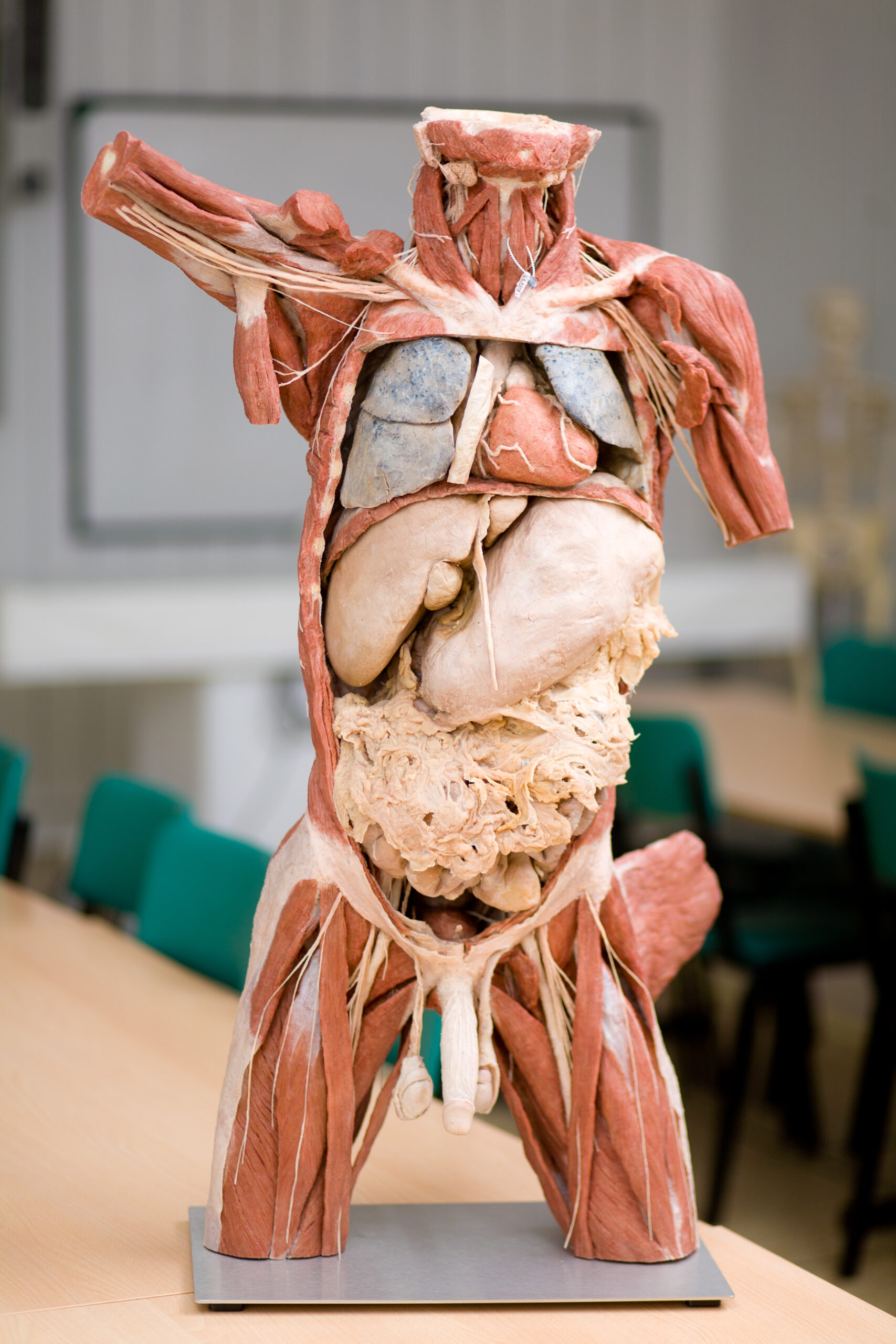
Plastinates of human bodies
- The plastination method was invented by German anatomist Gűnter von Hagens in 1977 to preserve body specimens for medical education. In the process, body fluids and fat are replaced by liquid plastics in the samples.
- After the bodies are positioned in realistic positions, they are hardened by gas, heat or light. The plastinates then show how our bodies move in everyday life and during sporting activities. Plastination has brought significant improvements to anatomical research and teaching through visual and tactile sensation and realism.
- Today, plastinates are already helping virtually all over the world in teaching anatomy and allowing students to gain unique experiences. All of Hagens’ anatomical specimens come from the bodies of donors who donated their bodies with official permission during their lifetimes, for the purpose of educating future generations.
- The donor programme has been running for almost 35 years. At the Faculty of Health Studies, plastinated human bodies and limbs help, for example, in the training of paramedics, physiotherapists or nurses. They enable them to study the human body literally down to the last blood vessel.


Did you know that…
Students in the Paramedic program solve approximately 60 model situations during their three years of study.
ambulance of the medical rescue service
- This facility is equipped with all the instruments, technology and furniture, including a stretcher with a very realistic, highly sophisticated model of the patient. Among the instruments are fully functional resuscitation machines, ECGs, oximeters, dosing devices, defibrillation trainers, etc.
- There is also the necessary medical material used in the treatment. The simulator is equipped with a camera system that allows to record the activities of the learners in the car and to project it on a large screen so that both the teacher and the students can watch the course of the rehearsed intervention.
Klára Gillnerová
Did you know that…
Throughout their studies, students of the Paramedic program complete 1320 hours of professional practice at medical workplaces?